Lipid nanoparticles engineered to specifically target pancreas in mouse model


Artistic representation of a lipid nanoparticle containing mRNA. Credit:iStock

Therapeutics that use mRNA—like some of the COVID-19 vaccines—have enormous potential for the prevention and treatment of many diseases. These therapeutics work by shuttling mRNA “instructions” into target cells, providing them with a blueprint to make specific proteins. These proteins could help tissues to regenerate, replace misfunctioning proteins, or prompt an immune response, providing a variety of different treatment strategies.

However, a therapeutic is only useful if it can reach its target. The mRNA is typically packaged inside a lipid nanoparticle, which keeps the delicate cargo intact until it reaches its final destination. As the field stands now, mRNA-filled lipid nanoparticles generally reach just a handful of cell types, such as immune cells and cells in the liver or spleen. Designing such lipid nanoparticles that can target hard-to-reach organs, such as the heart or pancreas, could revolutionize treatment options for a wide range of conditions.

In response to this need, researchers at Carnegie Mellon University are developing lipid nanoparticles that are designed to carry mRNA specifically to the pancreas. Their study in mice, recently published in Science Advances, could pave the way for novel therapies for intractable pancreatic diseases, such as diabetes and cancer.

“Lipid nanoparticles are essentially tiny spheres of fat, and fats have all kinds of chemical properties that can affect their ability to travel through the body and target specific organs,” explained Luisa Russell, Ph.D., a program director in the Division of Discovery Science & Technology at the National Institute of Biomedical Imaging and Bioengineering (NIBIB). “By optimizing these fat molecules and investigating alternative drug delivery routes, the study authors were able to design a lipid nanoparticle that can safely deliver mRNA to pancreatic tissue in mice.”

Current mRNA drug delivery routes include intramuscular injection (used in COVID-19 vaccines) and intravenous administration (used in some investigational cancer therapeutics). As a first step towards targeted delivery, the study authors wanted to know if a different administration route might help deliver the mRNA cargo directly to the pancreas. They investigated mRNA delivery via intraperitoneal injection, which involves injecting a drug directly into the fluid that surrounds the organs of the peritoneal cavity (including the kidneys, intestines, and pancreas).

“While intraperitoneal injection is not commonly used in humans, this type of administration is used clinically for some difficult-to-treat diseases, such as ovarian cancer,” said senior study author Kathryn Whitehead, Ph.D., a professor at Carnegie Mellon University. “With very serious pancreatic diseases, the benefits of intraperitoneal injection outweigh the risks.”

The researchers packaged mRNA instructions for firefly luciferase—a bioluminescent protein often used in research—into lipid nanoparticles, and then injected them into mice either intravenously or intraperitoneally. Using the glowing firefly luciferase to see where the mRNA had traveled, they found that intraperitoneal injection resulted in more abundant and more specific delivery to the pancreas compared with intravenous injection.

Next, the researchers began to optimize the composition of fat molecules that make up the nanoparticle. Different fats have unique chemical properties—such as size, electrical charge, and hydrophobicity—that can affect what happens to the nanoparticle once it enters the body. One type of fat molecule used is called a “helper lipid,” so named because it helps to stabilize the nanoparticle and improves its potency. The researchers wanted to know if changing the charge of the helper lipid might affect the targeting of the nanoparticle and direct it towards the pancreas. After trying a variety of different nanoparticle compositions, the researchers found a combination of lipids that improved pancreatic targeting in mice.

“Within the past couple of years, there’s been much more appreciation for how the lipids in nanoparticles can redirect mRNA delivery to different cells and organs,” said first study author Jilian Melamed, Ph.D., a postdoctoral researcher at the University of Pennsylvania. “The precise ways that lipid chemistry affects the potency and specificity of nanoparticles are still being uncovered, and we are still working to understand how individual lipid components influence overall mRNA delivery.”

When the authors investigated where exactly their optimized nanoparticles were going in the pancreas, they were surprised to discover that mRNA was most abundant in pancreatic islet cells, which comprise only 1%–2% of total pancreatic tissue. Pancreatic islet cells are responsible for producing hormones that control glucose in the blood (such as insulin). Such specific targeting could have potential downstream clinical applications.

“With further development, our research may lead to the creation of therapies for diabetes or certain types of pancreatic cancer,” said Whitehead. “These potential treatments, however, would require more preclinical research before advancing to clinical trials.”

Source/Imagesource

Read the Latest Genesis Nanotech Online: Therapeutic viruses help turbocharge the immune system against cancer


The illustration shows a cancer cell (center) surrounded by immune T-cells augmented with an oncolytic (cancer-fighting) virus. A new study describes how a combination of immunotherapy and virotherapy, using myxoma virus, provides new hope for patients with treatment resistant cancers. Credit: Jason Drees

The immune system has evolved to safeguard the body from a wildly diverse range of potential threats. Among these are bacterial diseases, including plague, cholera, diphtheria and Lyme disease, and viral contagions such as influenza, Ebola virus and SARS CoV-2.

Despite the impressive power of the immune system’s complex defense network, one type of threat is especially challenging to combat. This arises when the body’s own native cells turn rogue, leading to the phenomenon of cancer. Although the immune system often engages to try to rid the body of malignant cells, its efforts are frequently thwarted as the disease progresses unchecked.The illustration shows a cancer cell (center) surrounded by immune T-cells augmented with an oncolytic (cancer-fighting) virus. A new study describes how a combination of immunotherapy and virotherapy, using myxoma virus, provides new hope for patients with treatment resistant cancers. 

In new research appearing in the journal Cancer Cell, corresponding authors Grant McFadden, Masmudur Rahman and their colleagues propose a new line of attack that shows promise for treatment-resistant cancers.

The approach involves a combination of two methods that have each shown considerable success against some cancers. The study describes how oncolytic virotherapy, a technique using cancer-fighting viruses, can act in concert with existing immunotherapy techniques, boosting the immune capacity to effectively target and destroy cancer cells.

Oncolytic viruses represent an exciting new avenue of cancer therapy. Such viruses have the remarkable ability to hunt and terminate cancer cells while leaving healthy cells unharmed, as well as enhancing the immune system’s ability to recognize and terminate cancer cells.

One such virus, known as myxoma, is the focus of the current research and an area of expertise for the research group. The study shows that the use of T-cells infected with myxoma virus can induce a form of cancer cell death not previously observed.

Known as autosis, this form of cell destruction may be particularly useful against solid tumors that have proven treatment-resistant to various forms of cancer therapy, including immunotherapy alone.

“This work affirms the enormous potential of combining virotherapy with cell therapy to treat currently intractable cancers,” McFadden says.

McFadden directs the Biodesign Center for Immunotherapy, Vaccines and Virotherapy at Arizona State University.

Internal sentries

The immune system is composed of a range of specialized cells designed to patrol the body and respond to threats. The system is involved in a ceaseless arms race against pathogens, which evolve sophisticated techniques to attempt to outwit immune defenses, propagate in the body and cause disease. Cancer presents a unique challenge to the immune system as tumor cells often lack the identifying cell features that allow the immune system to attack them by distinguishing self from non-self.

Cancer cells can further short-circuit immune efforts to hunt and destroy them, through a range of evasive strategies. Researchers hope to help the immune system to overcome cancer’s notorious tactics of disguise, developing new experimental techniques belonging to a category known as adoptive cell therapy, or ACT.

Such methods often involve removing a collection of cancer-fighting white blood cells known as T-cells, modifying their seek-and-destroy capacities and reinjecting them in patients. Two forms of ACT immunotherapy are described in the new study: CAR T-cell therapy (CART) and T Cell Receptor Engineering (TCR). The basic idea in each case is the same: treating cancer with activated T lymphocytes extracted from the patient.

New method delivers one-two punch to tumor cells

The development of these therapies has been nothing short of revolutionary, and some cancer patients facing grim prospects have made remarkable recoveries following the use of immunotherapy. But techniques like CART and TCR nevertheless have their limitations and are often ineffective against advanced solid tumors. In such cases, cancer cells often manage to evade destruction by T-cells by downregulating or losing the surface antigens or MHC proteins that T-cells use to identify them. 

The new study highlights the ability of immunotherapy when it is coupled with virotherapy to break through the wall of cancer resistance, specifically using myxoma-equipped T-cells. The myxoma can target and kill cancer cells directly but more usefully can induce an unusual form of T-cell directed cell death known as autosis. This form of cell death augments two other forms of programmed cancer cell death induced by T-cells, known as apoptosis and pyroptosis. 

During myxoma-mediated autosis, cancerous cells in the vicinity of those targeted by the therapy are also destroyed in a process known as bystander killing. This effect can considerably enhance the dual therapy’s aggressive eradication of cancer cells, even in notoriously hard-to-treat solid tumors.

A combined myxoma-immunotherapy approach therefore holds the potential to turn so-called “cold tumors,” which fly under the immune system‘s radar, into “hot tumors” that immune cells can identify and destroy, allowing CAR T-cells or TCR cells to enter the tumor environment, proliferate and activate. 

“We are at the edge of discovering newer aspects of the myxoma virus and oncolytic virotherapy,” Rahman says. “In addition, these findings open the door for testing cancer-killing viruses with other cell-based cancer immunotherapies that can be used in cancer patients.” 

The ability to radically reengineer oncolytic viruses like myxoma to target a range of resistant cancers provides a new frontier for the treatment of this devastating disease

+ Explore further

New review highlights cancer-crushing viruses

More information: Ningbo Zheng et al, Induction of tumor cell autosis by myxoma virus-infected CAR-T and TCR-T cells to overcome primary and acquired resistance, Cancer Cell (2022). DOI: 10.1016/j.ccell.2022.08.001

Journal information: Cancer Cell 

Monash Biomedicine Develops New Approach for Bolstering T-Cells Ability to Fight Cancer


Credit: CC0 Public Domain

A collaborative study led by the Monash Biomedicine Discovery Institute (BDI) has discovered a new immune checkpoint that may be exploited for cancer therapy

The study shows that by inhibiting the protein tyrosine phosphatase PTP1B in T cells, the body’s immune response to cancer can be mobilized, helping to repress tumor growth.

T cells are an essential part of the body’s immune system, helping not only to kill invading pathogens, such as viruses but also cancer cells. However, this study has shown that the abundance of PTP1B in T cells that infiltrate tumors is increased, thereby restraining the ability of T cells to attack tumor cells and combat cancer. These findings have identified PTP1B as an intracellular brake, or checkpoint, reminiscent of the cell surface checkpoint PD-1—the blockade of which has revolutionized cancer therapy. 

The findings are published in the prestigious journal Cancer Discovery.

Using mice, scientists from Monash BDI, in conjunction with colleagues at the Peter MacCallum Cancer Center in Melbourne and Cold Spring Harbor Laboratory in New York, found that by inhibiting PTP1B, using an early-stage injectable drug candidate that has previously been shown to be safe and well-tolerated in humans, the cancer-fighting ability of T cells is enhanced, repressing tumor growth.

Remarkably, the authors showed that the inhibition of this intracellular checkpoint, PTP1B, can also enhance the response to a widely used cancer therapy that blocks the PD-1 checkpoint on the surface of T cells.

Senior author Professor Tony Tiganis says that although the blockade of PD-1 can be highly effective against many tumors, not all patients respond and the development of resistance is common. This is true even for immunotherapy-sensitive cancers, such as melanoma. Approaches that can enhance the effectiveness or extend the utility of PD-1 checkpoint blockade are highly sought after in the clinic.

“While more pre-clinical work is needed, our findings show that superior outcomes were achieved when we combined PTP1B inhibition with existing immunotherapies in mice,” said Professor Tiganis.

In addition, beyond enhancing the response to PD-1 blockade, the authors showed that the inhibition of PTP1B also significantly enhanced the effectiveness of cellular therapies using Chimeric Antigen Receptor (CAR) T cells.

CAR T cells are T cells derived from a patient’s blood that are modified in the lab so that they produce a man-made receptor to help them better identify tumor cells and then injected back into the patient. 

CAR T cells have been highly effective against some blood cancers; however, this success has not, as yet, been replicated in solid tumors. The authors demonstrate that the deletion or inhibition of PTP1B can dramatically enhance the ability of CAR T cells to attack solid tumors in mice, including breast cancer. 

“To advance this work, a key next step will be to further define the impact of PTP1B deletion in CAR T and conventional T cells in humans. There remains an urgent clinical need to identify and validate cellular targets to revive and sustain T cell responses in cancer,” said first author Dr. Florian Wiede.

Professor Tiganis and Dr. Wiede will also continue to collaborate with Cold Spring Harbor Laboratory and DepYmed Inc., a US-based company developing PTP1B inhibitors, to test in their preclinical models orally bioavailable PTP1B inhibitor drug candidates as novel checkpoint inhibitors. These findings could form the basis of future clinical trials.

Cancer continues to be a major cause of illness and death in Australia, accounting for 30 percent of all deaths in Australia in 2020. The AIHW cancer in Australia report estimates that around 185,000 cases of cancer will be diagnosed in 2031 and that between 2022 and 2031, a total of around 1.7 million cases of cancer will be diagnosed.

The full paper in Cancer Discoveryjournal is titled “PTP1B is an intracellular checkpoint that limits T cell and CAR T cell anti-tumor immunity.”

Increasing the capacity of the immune system to kill cancer cells


Graphical abstract. Credit: DOI: 10.1016/j.celrep.2021.110111

Awakening the immune system’s instinct for destroying cancer, using two molecules located on the surface of macrophages: that’s the promising avenue opening up from recent laboratory work of Dr. André Veillette.null

Director of the Molecular Oncology Research Unit of the Montreal Clinical Research Institute (IRCM) and a professor in the Department of Medicine at the Université de Montréal, Veillette recently published his findings in the journal Cell Reports.

His study unveils an innovative therapeutic way to treat cancer in line with the burgeoning field of precision medicine. For several years now, immunology and personalized medicine have brought new hope to physicians and patients in the fight against cancer.

These advanced therapies largely target cells of the immune system called T cells or T lymphocytes, whose role is to defend the body against harmful foreign agents such as viruses, bacteria and parasites, on the one hand, but also against cancer cells.

Among these “guardians of the body” are also macrophages, cells whose central role is to eliminate harmful agents by simply devouring them. There is a growing interest, among scientists and pharmaceutical companies, in targeting macrophages for therapeutic purposes.

In their lab, Dr. Veillette’s team discovered that macrophages are particularly good at destroying certain types of cancer cells. Even more, the team was able to greatly stimulate the appetite of these immune cells. In particular, they uncovered two molecules located on the surface of macrophages (CD11a and CD11c) which can be activated to increase their instinct to destroy macrophages.

In animal models and in human cell cultures in the lab, the stimulated macrophages turn into super-eaters of cancer cells.

“The ability to unleash the destructive power of macrophages is an important discovery that paves the way to some really exciting new possibilities in personalized medicine,” said Zhenghai Tang, co-first author of the study with Dominique Davidson. “In fact, added Davidson, “we help the body to protect itself better.”

This new use of the molecules to help the body cope better with cancer is an outgrowth of ongoing work in Dr. Veillette’s lab. He and his team have been studying the mechanisms that govern the functioning of the immune system for the past 30 years. In 2017, in a work published in the journal Nature, the team shed light on the SLAMF7 molecule, which also acts on the destructive capacity of macrophages.

“The more we know about the functioning of the immune system, the more we will be able to find effective and less toxic therapeutic solutions to fight diseases,” said Veillette. “Immune cells like macrophages are gaining a lot of interest in immunology research today, but also in the pharmaceutical industry, because this is truly the future of medicine for many deadly diseases.”

He added: “For our part, the next step will be to establish to what extent the molecules CD11a and CD11c can be used as biomarkers to identify patients who are most likely to respond to this type of therapy.”

Cancer chemotherapy drug reverses Alzheimer’s symptoms in mice – Read More at GenesisNanotech Online


GenesisNanotech – “Great Things From Small Thing”

Read GenesisNanotech Online: Articles Like: “Cancer chemotherapy drug reverses Alzheimer’s symptoms in mice” (Link) https://medicalxpress.com/news/2021-10-cancer-chemotherapy-drug-reverses-alzheimer.html

And … “Tiny bubbles can be future treatment for inflammation”

Scientists hope that tiny sacs of material excreted by cells—so-called extracellular vesicles—can be used to deliver drugs inside the body. (Link) https://medicalxpress.com/news/2021-10-tiny-future-treatment-inflammation.html

+More … Read The Latest Full Edition Here:

https://paper.li/GenesisNanoTech/1354215819#/

Biocompatible Thin Film: Heating Tissue with Surgical Precision to KILL CANCER


Human cells are vulnerable to intense heat and die rapidly above 42.5 °C. This property is utilized to treat cancer through a method called “thermotherapy” (also “hyperthermia”). The treatment has a long history, with even the ancient Greek physician Hippocrates being reported to use heat to eliminate cancer.

When tumor tissue is heated, the surrounding normal cells are also exposed to the heat. Although the blood vessels in normal tissue can dilate to release heat, the blood vessels in a tumor cannot, so only cancer cells reach a high temperature. It has also been reported that combining cancer thermotherapy (that works in this way) with radiation therapy and chemotherapy enhances the effect.

Although thermotherapy is promising as a cancer treatment, current methods require large devices for emitting radio waves or microwaves, so only a limited number of facilities are able to provide the treatment.

Applying polymeric thin film in medical treatment

Wearing a watch or other device on the body can cause skin irritation due to the mismatch in softness. Soft materials such as rubber can be used to avoid the mismatch, but this in turn leads to durability issues. To resolve this, Fujie Laboratory is developing a highly flexible polymeric thin film. Making the polymeric thin film thinner also makes it softer, allowing for the creation of flexible, comfortable devices (Advanced Functional Materials, “Flexible Induction Heater Based on the Polymeric Thin Film for Local Thermotherapy”).

Using an inkjet printer with conductive ink to draw a circuit on the polymeric thin film, it is possible to create a device that emits light and energy locally, with promising applications in medical treatment.

Local thermal device based on induction heating

The prototype was made based on the concept that when a polymeric thin film is attached to tumor tissue in vivo, and an alternating magnetic field is applied from outside the body, the thin film generates heat by the same principle as an induction cooktop.

Since the polymeric thin film is placed in vivo, the team decided to make it with biocompatible polylactic acid and use gold nano ink for inkjet printing. The gold nano ink needed to be heated to 250 °C to remove the stabilizer and induce conductivity. So it was printed on polyimide film made of a polymeric material that can withstand high temperature treatment.

After treatment at 250 °C, it was transferred to a polylactic acid thin film. The thickness of the thin film is only 7 µm, but it has the strength and flexibility to withstand handling with forceps used in endoscopic surgery.

The research team attached the device to the surface of an animal’s liver and applied a magnetic field. One minute of electricity raised the surface temperature by about 7 °C, and 5 minutes raised the temperature by about 8 °C. The liver used in the experiment was normal tissue, and a pathological examination afterwards revealed no burns or other damage.

Future prospects — Development of biocompatible medical devices

The polymeric thin film heating device can be sent non-invasively to tumor tissue using an endoscope. And by applying a magnetic field from outside the body, it is possible to heat the tumor tissue without the need for large equipment. This could likely lead to cancer thermotherapy becoming more widespread.

STINGing Tumors With Nanoparticles – Boosting the Body’s Innate Immune System to Fight Cancer


Synthetic-Polymer-Activates-STING-Proteins-777x777

This artist’s rendering shows a synthetic polymer (purple) that activates STING proteins (yellow and green motifs) for cancer immunotherapy. Credit: Shenyang Zhiyan Science and Technology Co. Ltd.

By  

New immunotherapy drug activates the body’s innate immune system to fight cancer.

A new nanoparticle-based drug can boost the body’s innate immune system and make it more effective at fighting off tumors, researchers at UT Southwestern have shown. Their study, published in Nature Biomedical Engineering, is the first to successfully target the immune molecule STING with nanoparticles about one millionth the size of a soccer ball that can switch on/off immune activity in response to their physiological environment.

“Activating STING by these nanoparticles is like exerting perpetual pressure on the accelerator to ramp up the natural innate immune response to a tumor,” says study leader Jinming Gao, Ph.D., a professor in UT Southwestern’s Harold C. Simmons Comprehensive Cancer Center and a professor of otolaryngology – head and neck surgery, pharmacology, and cell biology.

For more than a decade, researchers and pharmaceutical companies have been racing to develop drugs that target STING, which stands for “stimulator of interferon genes.” The STING protein, discovered in 2008, helps mediate the body’s innate immune system — the collection of immune molecules that act as first responders when a foreign agent circulates in the body, including cancer DNA. Research has suggested that activating STING can make the innate immune system more powerful at fighting tumors or infections. However, results from earlier clinical trials involving first-generation compounds targeting STING for activation failed to demonstrate an impressive clinical effect.


“A major limitation of conventional small molecule drugs is that after injection into tumors, they are washed out from the tumor site by blood perfusion, which can reduce antitumor efficacy while causing systemic toxicities,” explains Gao.

Jinming Gao

Jinming Gao, Ph.D. Credit: UT Southwestern Medical Center

Gao and his colleagues at UTSW discovered another approach that is different from the earlier or first-generation STING agonist approaches that utilize synthetic cyclic dinucleotide to activate STING in the body. Gao and his team aimed to design a polymer — a manmade macromolecule that can self-assemble into nanoparticles — to effectively deliver cyclic GMP-AMP (cGAMP), a natural small molecule activator of STING, to the protein target. But one polymer they synthesized, PC7A, produced an unexpected and novel effect: It activated STING even without cGAMP. The group reported the initial results in 2017, not knowing at the time exactly how PC7A worked; the polymer didn’t resemble any other drugs that activated STING.

In the new paper, Gao’s team showed that PC7A binds to a different site on the STING molecule from known drugs. Moreover, its effect on the STING protein is different. While existing drugs activate the protein over the course of about six hours, PC7A forms polyvalent condensates with STING for over 48 hours, causing a more sustained effect on STING. This longer innate immune activation, they showed, leads to a more effective T cell response against multiple solid tumors. Mice survived longer and had slower tumor growth when they received a combination of PC7A and cGAMP, the researchers found.

The polymer also has other advantages. When circulating in the bloodstream, the polymers are present as small round nanoparticles that do not bind to STING. It’s only when those nanoparticles enter immune cells that they separate, attach to STING, and activate the immune response. That means that PC7A might be less likely to cause side effects throughout the body than other STING-targeting drugs, says Gao, although clinical trials will be needed to prove that.

Because PC7A binds to a different site of the STING molecule, the compound might work in patients for whom typical STING-targeting drugs do not. Up to 20 percent of people have inherited a slightly different gene for STING; the variant makes the STING protein resistant to several cyclic dinucleotide drugs. Gao and his team demonstrated that PC7A can still activate cells that express these STING variants.

“There’s been a lot of excitement about therapies that target STING and the potential role these compounds could play in expanding the benefits of immunotherapies for cancer patients,” says Gao. “We believe that our new nanotechnology approach offers a way to activate STING without some of the limitations we’ve seen with earlier STING agonist drugs in development.”

Reference: “Polycarbonate-based ultra-pH sensitive nanoparticles improve therapeutic window” by Xu Wang, Jonathan Wilhelm, Wei Li, Suxin Li, Zhaohui Wang, Gang Huang, Jian Wang, Houliang Tang, Sina Khorsandi, Zhichen Sun, Bret Evers and Jinming Gao, 17 November 2020, Nature Communications.
DOI: 10.1038/s41467-020-19651-7

Other UTSW researchers who contributed to this study were Suxin Li, Min Luo, Zhaohui Wang, Qiang Feng, Jonathan Wilhelm, Xu Wang, Wei Li, Jian Wang, Agnieszka Cholka, Yang-xin Fu, Baran Sumer, and Hongtao Yu.

This research was supported by funds from the National Institutes of Health (U54 CA244719) and Mendelson-Young Endowment for Cancer Therapeutics.

Gao holds the Elaine Dewey Sammons Distinguished Chair in Cancer Research, in honor of Eugene P. Frenkel, M.D. at UTSW.


Getting more Cancer-Fighting Nanoparticles to where they are Needed: University of Toronto


2-chemotherapyCredit: CC0 Public Domain

University of Toronto Engineering researchers have discovered a dose threshold that greatly increases the delivery of cancer-fighting drugs into a tumor.

Determining this threshold provides a potentially universal method for gauging nanoparticle dosage and could help advance a new generation of cancer therapy, imaging and diagnostics.

“It’s a very simple solution, adjusting the dosage, but the results are very powerful,” says MD/Ph.D. candidate Ben Ouyang, who led the research under the supervision of Professor Warren Chan.

Their findings were published today in Nature Materials, providing solutions to a drug- problem previously raised by Chan and researchers four years ago in Nature Reviews Materials.

Nanotechnology carriers are used to deliver drugs to cancer sites, which in turn can help a patient’s response to treatment and reduce , such as hair loss and vomiting. However, in practice, few injected particles reach the tumor site.

In the Nature Reviews Materials paper, the team surveyed literature from the past decade and found that on median, only 0.7 percent of the chemotherapeutic nanoparticles make it into a targeted tumor.

“The promise of emerging therapeutics is dependent upon our ability to deliver them to the target site,” explains Chan. “We have discovered a new principle of enhancing the delivery process. This could be important for nanotechnology, genome editors, immunotherapy, and other technologies.”

Chan’s team saw the liver, which filters the blood, as the biggest barrier to nanoparticle drug delivery. They hypothesized that the liver would have an uptake rate threshold—in other words, once the organ becomes saturated with nanoparticles, it wouldn’t be able to keep up with . Their solution was to manipulate the dose to overwhelm the organ’s filtering Kupffer cells, which line the liver channels.

The researchers discovered that injecting a baseline of 1 trillion nanoparticles in mice, in vivo, was enough to overwhelm the cells so that they couldn’t take up particles quick enough to keep up with the increased doses. The result is a 12 percent delivery efficiency to the tumor.

“There’s still lots of work to do to increase the 12 percent but it’s a big step from 0.7,” says Ouyang. The researchers also extensively tested whether overwhelming Kupffer cells led to any risk of toxicity in the liver, heart or blood.

“We tested gold, silica, and liposomes,” says Ouyang. “In all of our studies, no matter how high we pushed the dosage, we never saw any signs of toxicity.”

The team used this threshold principle to improve the effectiveness of a clinically used and chemotherapy-loaded nanoparticle called Caelyx. Their strategy shrank tumors 60 percent more when compared to Caelyx on its own at a set dose of the chemotherapy drug, doxorubicin.

Because the researchers’ solution is a simple one, they hope to see the threshold having positive implications in even current nanoparticle-dosing conventions for human clinical trials. They calculate that the human threshold would be about 1.5 quadrillion nanoparticles.

“There’s a simplicity to this method and reveals that we don’t have to redesign the  to improve delivery,” says Chan. “This could overcome a major delivery problem.”


Explore further

Researchers find more precise way to target tumours with anti-cancer drugs

Tiny Nanoparticles Offer Large Potential for Brain Cancer Treatment


tiny brain nanoparticles 1-tinynanopartFor patients with malignant brain tumors, the prognosis remains dismal. With the most aggressive treatments available, patients are usually only expected to live about 14 months after a diagnosis
This is because, chemotherapy, the most common form of treatment for cancer, is uniquely challenging for   patients. The delicate organ in our skulls is protected by a network of vessels and tissue called the blood-brain barrier that keeps most foreign substances out. Furthermore,  can cause significant damage to the rest of the body if they are not able to target the tumor in a pharmacologically significant dose.

These challenges have plagued scientists for years, but a team of researchers for Yale School of Medicine and Beijing Normal University just published a breakthrough study detailing a new method that offers a promise at treatment. The solution? Nanoparticles.

Nanoparticles, particles that are smaller than wavelengths of visible light and can only be seen under a special microscope, have the potential to pass through the blood-brain barrier. They can also carry drugs to targeted areas of the body, reducing the side effects on the rest of the body. But previous nanoparticles were very complex and not very efficient in penetrating in the brain.

This most recent paper, published in Nature Biomedical Engineering on March 30, 2020, describes a small carbon nanoparticle engineered by the two labs that could both deliver chemotherapy drugs across the blood-brain barrier and mark tumor cells with fluorescence in mice. What’s more, this nanoparticle is incredibly simple—made up of only one single compound.

“The major problems we’ve solved is to improve the delivery efficiency and specificity of nanoparticles,” says Jiangbing Zhou, Ph.D., associate Professor of Neurosurgery and of Biomedical Engineering at Yale School of Medicine. “We created nanoparticles like building a missile. There’s usually a GPS on every missile to guide it into a specific location and we’re able to guide particles to penetrate the brain and find tumors.”

The GPS-like targeting occurs because the nanoparticles engineered to be recognized by a molecule called LAT1, which is present in the blood-brain  as well as many tumors, but not in most other normal organs. As a result, chemotherapy drugs can be loaded on the dots and target tumors while barely affecting the rest of the body. The nanoparticles gain entry to the brain because they’ve been engineered to look like amino acids, which are allowed past the  as nutrients.

The nanoparticles have wider implications than  delivery. They can be stimulated to emit a fluorescence, which helps surgeons locate tumor to remove with greater accuracy.

Still, there’s a long road ahead before this research can be applied in a clinical setting, says Dr. Zhou. “It takes a long time before the technology can be translated into clinical applications,” he says. “But this finding suggests a new direction for developing  for drug delivery to the brain by targeting LAT1 molecules.”


Explore further

Improving drug delivery for brain tumor treatment


More information: Shuhua Li et al. Targeted tumour theranostics in mice via carbon quantum dots structurally mimicking large amino acids, Nature Biomedical Engineering (2020). DOI: 10.1038/s41551-020-0540-y

Journal information: Nature Biomedical Engineering

Unmasking a hidden killer: Successfully detecting cancer in blood of patients undergoing treatment


unmaskingahi
Dr Yuling Wang. Credit: CNBP

Pancreatic cancer is one of the most lethal cancers, but difficult to diagnose: few sufferers have symptoms until the cancer has become large or already spread to other organs. Even then, symptoms can be vague and easily misconstrued as more common conditions.

This is why Dr. Yuling Wang is so excited by results of a trial completed in late 2019, which—using plasmonic nanoparticles developed by the Centre for Nanoscale BioPhotonics (CNBP)—successfully detected signs of the  in  of patients undergoing treatment. The paper was recently published in the journal American Chemical Society—Sensors.

“The test gave a very high signal in the blood for late-stage or very serious tumors, where other techniques cannot detect anything,” said Dr. Wang, an associate investigator at the Centre’s Macquarie University node in Sydney, in work led by Prof Nicolle Packer. “We need to test many more patient samples to validate the approach, but the strength of the signal was very encouraging.”

They did this by developing a method, using surface-enhanced Raman spectroscopy nanotags, that simultaneously detects three known  cancer biomarkers in blood. Known as extracellular vesicles, or EVs, they contain DNA and proteins for cell-to-cell communication and are shed from pancreatic cancer cells into surrounding body fluids. The CNBP method zeros in on three: Glypican-1, epithelial cell adhesion molecules and CD44V6.

Unmasking a hidden killer

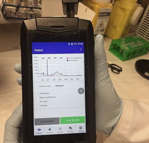
Non-invasive screening of cancer biomarkers from blood with handheld Raman reader. Credit: CNBP

For the experiment, biopsies of healthy donors were provided alongside those of known sufferers of pancreatic cancer, in double-blind tests where the researchers did not know which was which. Nevertheless, the blood of sufferers was easily identified. The technique was so sensitive it could spot EVs as small as 113 nanometres in diameter—or less than 1% the width of a human hair—in every millilitre of blood.

The pancreas is part of the digestive system, secreting insulin into the bloodstream to regulate the body’s sugar level as well as important enzymes and hormones into the  to help break down food. Pancreatic cancer is the fifth biggest cancer killer in Australia and has a 5-year survival rate of 8.7%. More than 3000 Australians are diagnosed annually, and surgery to remove the cancer is a long and complex process, requiring long hospital stays.

Because existing blood tests for the protein biomarkers of pancreatic cancer are unreliable, imaging with endoscopic ultrasound or MRI scans is necessary. Even then, anomalies can only be confirmed with a biopsy of the organ, which is invasive and ultimately relies on a trained pathologist to recognize signs of the cancer under a microscope. As a result, there’s some subjectivity involved and cancer can be present but still be missed.

“Our approach is non-invasive—we don’t need to take tissue from the patient, we just use a  to test blood for targeted biomarkers, which gives a very quick result,” Dr. Wang said. It may also help provide earlier diagnosis of the cancer.

While the work is a proof-of-concept, it was also able to detect colorectal and bladder cancer biomarkers—although not as clearly as those for . Nevertheless, the results are so encouraging that a commercial partner has committed funding to CNBP so it can develop a handheld spectrometer for cancer biomarkers in blood.


Explore further

UK urine test that can detect early-stage pancreatic cancer starts clinical study